Dr. Jan Philipp Schneider and colleagues aimed to design a 3D reconstructions of type 1 human alveolar epithelial cells (AE1 cells) to gain a deeper understanding of their complex structure.
In close collaboration with MHH's Central Research Facility for Imaging of the DZL, a picture block of more than 2000 images from a human lung sample was obtained for this purpose using a so-called Serial Block-Face Scanning Electron Microscope. This microscope automatically removes thin sections of the specially prepared sample with a diamond knife. Between the individual cutting processes, the newly developed surface of the sample is scanned with an electron beam and, thus, recorded with high resolution. This creates a stack of images that can be used for the reconstructions. The scientists at BREATH were then able to create 3D models of three complete AE1 cells by manual segmentation (i.e. manual tracking of the cells on the individual images) and gain new insights into the 3D structure of the alveolar region.
The paper, which has now been named "Paper of the Month" by the Anatomical Society, has been published in the highly cited American Journal of Respiratory and Critical Care Medicine. A related video showing the EM record and models and their origins can be viewed on the American Thoracic Society's YouTube® channel.
You can find the original publication here.
Text: BREATH / CD
Picture: Institute for Functional and Applied Anatomy, MHH
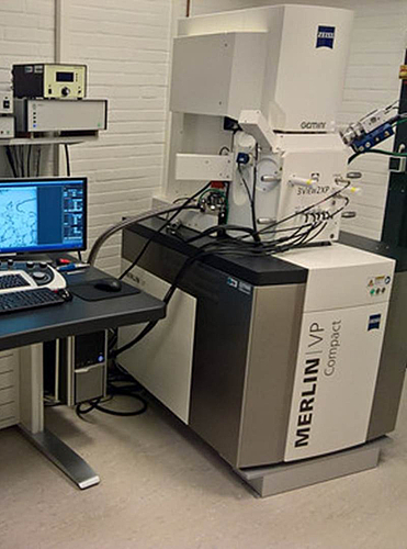
[Translate to English:] Das verwendete Rasterelektronenmikroskop zur seriellen Abbildung der Blockoberfläche für die 3D-Rekonstruktion (Serial Block Face SEM)